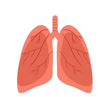
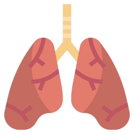
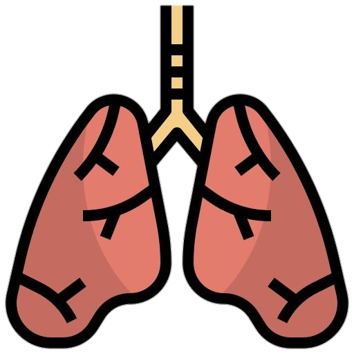
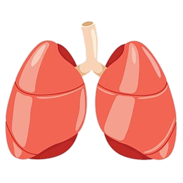
This gallery shows 18+ high-quality and best-resolution Lungs PNG Images, Vectors, Stickers, logos, Icons, and Clipart Pictures with transparent backgrounds. Free download all these Lungs PNG images for graphic design, projects, presentations, web design, editing, and other works.
Lungs PNG Images:

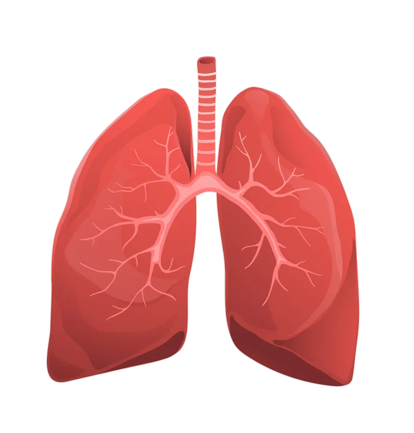
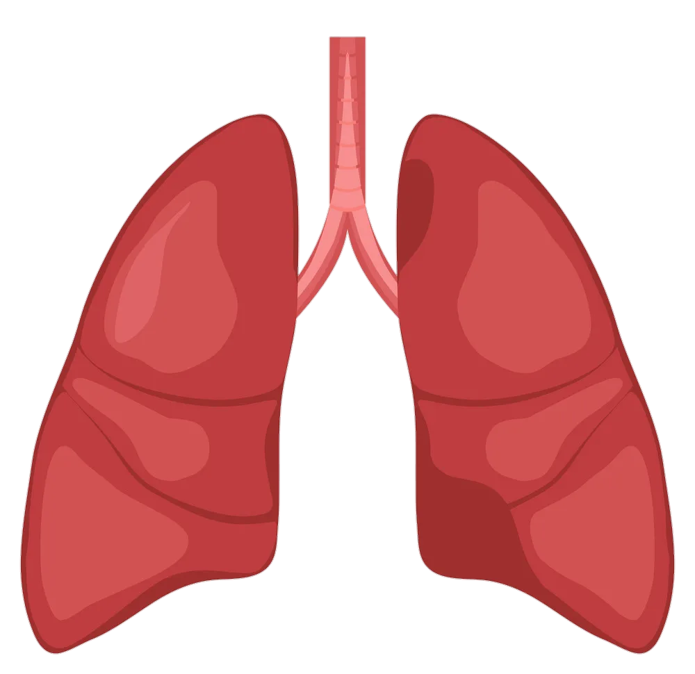
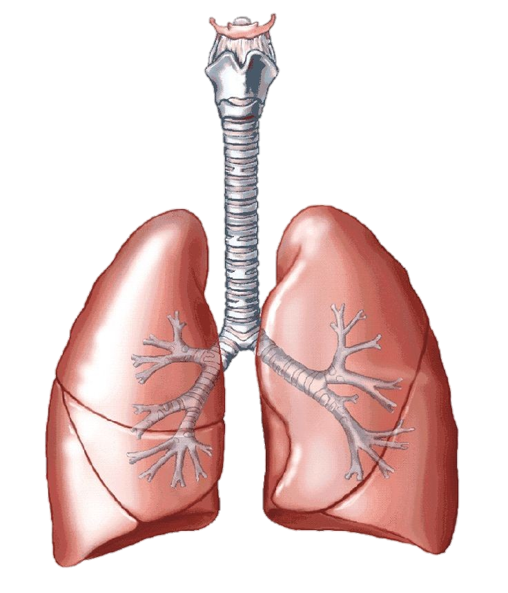
The human lungs are the primary organs responsible for respiration, taking in oxygen, and expelling carbon dioxide. They are part of the respiratory system, along with the airways and muscles involved in breathing. The lungs are located in the thoracic cavity, on either side of the heart, and are protected by the ribcage.
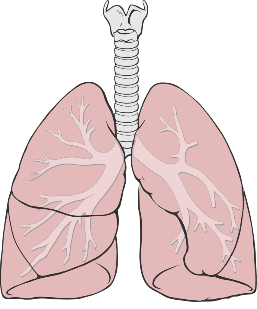
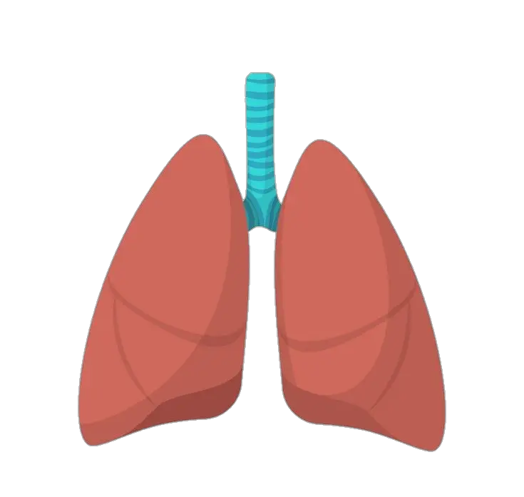
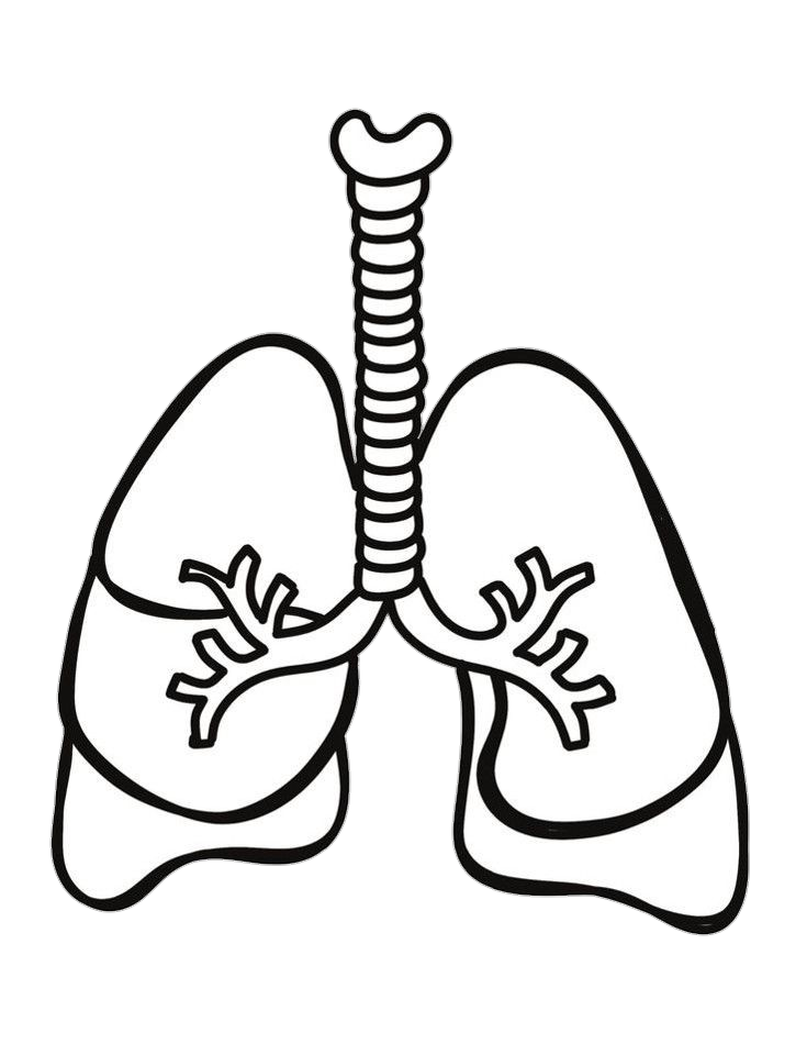
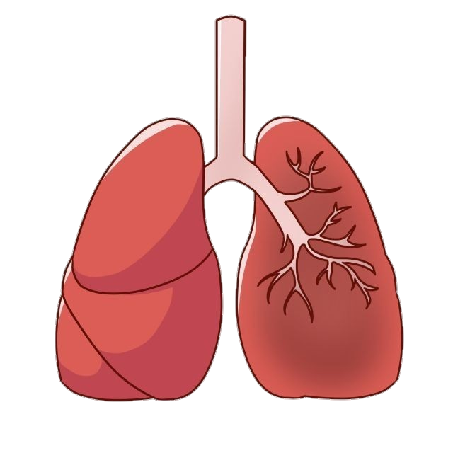
Each lung is a conical-shaped organ divided into lobes. The right lung has three lobes (upper, middle, and lower), while the left has two lobes (upper and lower). This asymmetry is due to the heart’s presence on the chest’s left side.
The lungs are composed of spongy, elastic tissue surrounded by a double-layered membrane called the pleura. The outer layer, the parietal pleura, lines the chest wall, while the inner layer, called the visceral pleura, covers the lungs. The space between these two layers is known as the pleural cavity and contains a small amount of fluid, which helps reduce friction during breathing movements.
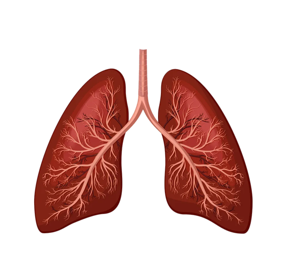
The lungs’ main function is to facilitate the exchange of gases between the atmosphere and the bloodstream. When we inhale, air enters the body through the nasal passages or mouth and travels down the trachea (windpipe). The trachea branches into two tubes called bronchi, which further divide into smaller branches known as bronchioles. These bronchioles eventually end in tiny air sacs called alveoli.
The alveoli are the site of gas exchange. They are surrounded by an extensive network of blood vessels called capillaries. Oxygen from the inhaled air diffuses across the thin walls of the alveoli into the capillaries, where it binds to red blood cells. At the same time, carbon dioxide, a waste product of metabolism, passes from the capillaries into the alveoli and is expelled when we exhale.
The lungs also play a role in regulating the body’s acid-base balance. By controlling carbon dioxide levels in the blood, the lungs help maintain the pH within a narrow range, ensuring proper physiological function.
Breathing, or ventilation, moves air in and out of the lungs. It is controlled by the respiratory center in the brain, which sends signals to the muscles involved in respiration. When we inhale, the diaphragm (a dome-shaped muscle located below the lungs) contracts and moves downward, while the muscles between the ribs contract and lift the ribcage, these actions increase the volume of the chest cavity, causing a decrease in pressure within the lungs. As a result, air flows in from the higher-pressure environment outside the body, filling the lungs. During exhalation, the muscles relax, the chest cavity decreases volume, and the air is expelled from the lungs.